Today, the photographs and Wilson’s interpretation seem to be convincing. Some biologists, however, maintained that the rays and spindles were actually artefacts of fixing, staining, and visualizing the cells. Understanding cell division is essential to understanding embryonic development. When cell division occurs, both daughter cells receive the same complement of genetic material in the chromosomes parcelled out to both cells. In the parent cell certain proteins and other important molecules may have been unequally distributed, and this would mean that after division, the daughter cells would have unequal proportions of these molecules. This would potentially lead them towards different fates; one sister committed to be one type of cell perhaps, the other sister committed to be another. Scientists in the early twentieth-century such as Wilson and Thomas Hunt Morgan (1866-1945) realized this.
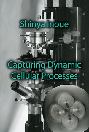
To understand this in broad terms was one thing, to demonstrate it was quite another. To demonstrate how the mechanics of cell division worked, biologists would need to develop new ways to visualize structures in live cells and demonstrate their nature and function. Among other technical and scientific achievements, Shinya Inoué was instrumental in demonstrating the role and operation of the spindle in cell division. He accomplished this by finding ways of visualizing structures in live cells, using the properties of polarized light to develop new optical microscopes. Inoué and his colleagues used polarized light to reveal the structures in the cell in real-time, and helped to produce the dynamic view of the cell that we have today.
- Shinya Inoué, Pathways of a Cell Biologist. Through Yet Another Eye. Springer: 2016
- Shinya Inoué, Collected Works Of Shinya Inoué: Microscopes, Living Cells, and Dynamic Molecules (With DVD-ROM). World Scientific Publishing Company; Har/Dvdr edition (July 18, 2008).

