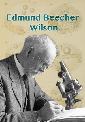
With a quick glance at Wilson’s figures in Cell-Lineage, it is easy to appreciate how carefully drawn and beautifully colored they are. A closer look reveals that they are not only the culmination of years of labor and careful observation, but they are also the end result of a long chain of images produced through various methods and mediums. Hundreds of initial sketches and observations were compiled into ninety-two drawings, which were then copied by a lithographer onto eight lithography plates. The prints of those plates are the final images bound in the publication.
Like most of his contemporaries, Wilson used a camera lucida to produce most of the drawings for his figures. Used widely throughout the nineteenth and twentieth centuries by both scientists and artists, a camera lucida allows for a drawing surface to be seen simultaneously with the specimen of interest through the eyepiece of the microscope. Through a series of angled mirrors, the drawing surface and pen or pencil is superposed onto the specimen such that it can be accurately traced. Although none of Wilson’s original sketches have survived, it is likely that he made hundreds of camera lucida sketches as he was sitting at the microscope observing his embryos.
Wilson’s decision to use a camera lucida reflects his opinion that scientific claims could only be based on empirical observation (Maienschein 1991). Because specimens can essentially be traced, using a camera lucida allowed Wilson to make his figures and observations as accurate as possible and to ensure that his claims were based on phenomena he actually saw. Looking at the figures in Cell-Lineage, it is clear that they are depictions of actual specimens observed, not idealized forms. This is demonstrated by the fact that Wilson includes several images of the same stage or phenomenon to show the variations he observed across specimens (e.g. Figs 63 and 64). In such cases where variations presented themselves, Wilson did not attempt to make generalizations to unify the individual specimens. Rather, he presented exactly what he observed even if he did not yet have a conclusion about the reason for the variation.

Today, what is seen through a microscope can be captured instantly through attached cameras. Although the process of producing an image is more immediate, taking less time and labor, the same decisions about how to use those images must still be made. Like Wilson with his camera lucida sketches, biologists today often take hundreds of photographs of what they see through the microscope. Those images must then be sorted through, compiled, and sometimes even modified to produce publishable figures. Like Wilson, biologists today must decide how to interpret the images, how to account for variability between specimens, and how and to what extent photographs can be altered. Thus, no matter what kind of image is used, how that image is created, used, or manipulated to show results or craft an argument is of primary concern to every biologist.
Early Life and Education
- Allen, Garland E. “Wilson, Edmund Beecher.” Dictionary of Scientific Biography 14: 423–6.
- Maienschein, Jane, "Edmund Beecher Wilson (1856-1939)". Embryo Project Encyclopedia (2013-08-05). ISSN: 1940-5030 http://embryo.asu.edu/handle/10776/6044. embryo.asu.edu/pages/edmund-beecher-wilson-1856-1939.
- Morgan, Thomas Hunt. 1941. “Biographical Memoir of Edmund Beecher Wilson, 1856–1939,” National Academy of Sciences Biographical Memoirs 21: 315–42.
The Johns Hopkins University
- Moore, John A. 1987. "Edmund Beecher Wilson, Class of '81". American Zoologist 27(3): 785-796.
The Stazione Zoologica in Naples and Music
- Groeben, Christiane. 1985. "Anton Dohrn--The Statesman of Darwinism: To commemorate the 75th anniversary of the death of Anton Dohrn". Biological Bulletin 168: 4-25. www.biolbull.org/content/168/3S/4.full.pdf+html.
- Maienschein, Jane. 1985. "First impressions: American biologists at Naples". Biological Bulletin 168: 187-191. www.biolbull.org/content/168/3S/187.full.pdf+html.
Cell Lineage
- Fischer, Antje HL et al. (2012). “The normal development of Platynereis dumerilii (Nereididae, Annelida).” Frontiers in Zoology 7(31): 1-39.
- Gilbert, Scott. (2014). Developmental Biology, 10th edition. Sunderland, MA: Sinauer Associates Inc Publishers.
- MacCord, Kate, "Germ Layers". Embryo Project Encyclopedia (2013-09-17). ISSN: 1940-5030 http://embryo.asu.edu/handle/10776/6273.
- Maienschein, Jane. (1991). Transforming Traditions in American Biology, 1880-1915. Baltimore, MD: Johns Hopkins University Press.
- Pearse, Vicki, John Pearse, Mildred Buchsbaum & Ralph Buchsbaum. (1987). Living Invertebrates. Pacific Grove, CA: The Boxwood Press.
- Seaver, Elaine C. (2014). “Variation in spiralian development: insights from polychaetes.” Int. J. Dev. Biol 58: 457-467.
- Sedgwick, William T. & Edmund B. Wilson. (1895). An Introduction to General Biology. New York: H. Holt.
- Wilson, Edmund B. (1892). “The Cell-Lineage of Nereis: A Contribution to the Cytogeny of the Annelid Body.” Journal of Morphology 6(3): 361-481.

