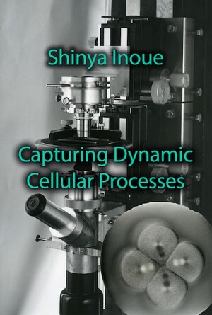
To investigate cell division and its consequence, scientists focused on ways of identifying parts of the cell, to find out how they acted and moved before, during, and after cell division. This therefore coincided with the other line of evidence about cells, through direct observation of them. As most cells are not visible to the naked eye, using a microscope was, and is, essential. In the seventeenth-century, Robert Hooke and Antonie van Leeuwenhoek were able to use their microscopes to observe the fine structure of insects, to identify cells, and to discover tiny living creatures including bacteria and protozoa.
Despite these technical and scientific achievements, at the beginning of the nineteenth-century microscopes were of limited use to scientists trying to observe early embryos, and in particular trying to observe cells. One of the problems that microscopes suffered from is known as chromatic aberration. Light traveling through the glass of the main lens, or objective, and through the glass of the eyepiece, refracts, or changes direction. Different wavelengths of light, corresponding to different colors, refract differently. When scientists tried to view objects at higher magnifications, this effect would prevent them from viewing it. Throughout the nineteenth-century, new techniques and designs of microscopes solved many problems like chromatic aberration. The optical microscope therefore became a valuable instrument for biologists in the latter half of the century.
Scientists also developed techniques to preserve and fix samples, to cut very thin slices using a device called a microtome, and stain whole cells or parts of them. They picked organisms that would help them in this task of visualizing cells and their innards. For example, many marine creatures such as sea urchins have transparent eggs, which enable scientists to observe early divisions of the egg, a process called cleavage or segmentation. The transparency of many marine creatures is one of the advantages that gave impetus to the establishment of research stations by the sea like the Marine Biological Laboratory at Woods Hole, and the Stazione Zoologica in Naples. In a relationship pioneered by its founder, Anton Dohrn (1840-1909), researchers at the Naples station had the discounted use of the latest equipment from the famous Carl Zeiss works in Germany, in exchange for the researchers making suggestions about improvements to the instruments.
- Shinya Inoué, Pathways of a Cell Biologist. Through Yet Another Eye. Springer: 2016
- Shinya Inoué, Collected Works Of Shinya Inoué: Microscopes, Living Cells, and Dynamic Molecules (With DVD-ROM). World Scientific Publishing Company; Har/Dvdr edition (July 18, 2008).

