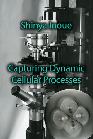
Designers of polarizing microscopes add extra components to the traditional setup of an optical microscope: a polarizer between the condenser and the stage, and an analyzer between the objective and the eyepiece. Both the polarizer and the analyzer are filters that polarize light. Microscopists arrange the polarizer and analyzer so that they are at right-angles to each other, so when non-polarizing materials are placed between them on the stage, no light makes it through to the eyepiece.
When Inoué set out to construct his microscope in the conditions of post-war Japan he could not just order them, or have them made in an instrument shop. He needed to find the components for himself. For a light source, he bought a mercury arc lamp from a surplus store. He obtained a prism from a professor in the Physics Department at Toh-Dai and got a Dichrome analyser from Katy’s microscope. Inoué wanted to follow the progress and changes of the mitotic spindle through the process of cleavage in the sand-dollar, Clypeaster japonica, a species with transparent eggs. Whether the spindle appears dark or light depends entirely on the orientation of the spindle; the important thing was that the spindle was distinguished from the background. Unfortunately, though he could see the birefringent spindles just over 30 minutes after fertilization, when the egg began to enter cleavage the spindles would no longer be visible. He therefore needed to improve the sensitivity of his polarizing microscope.
It took Inoué over a month to diagnose why his initial attempts at improving the microscope failed, and after this he decided that he needed to add an extra component, known as a compensator, which makes parts of the specimen with the same orientation of birefringence appear much brighter, with others appearing darker. After initial frustration, and then consternation at his attempted solutions making the situation worse, Inoué had found a way to increase the sensitivity of his polarizing microscope to the birefringence of the mitotic spindle.
In microscopy, scientists aim to improve the resolution and the contrast of the image. Inoué was not able to increase the resolution, as optical microscopy had already reached the maximum possible resolution by this time. He was able to increase the contrast, and his first microscope achieved this, as did his subsequent microscopes and techniques. Importantly, he was able to do this while keeping cells alive, and in conditions which kept them dividing. This enabled Inoué and Katy to view the changes in the spindle and asters over several cleavage cycles. By keeping cells alive, and in a term used by Katy, ‘happy’, they were able to observe the dynamic changes occurring in the living cell.
Inoué only created what was later named the ‘Shinya-scope’ when the Emperor of Japan visited the laboratory. Until the visit, Shinya had simply stood the components of the microscope on piles of books. For the visit, Shinya attempted to create a more stable configuration, and went into the woods to find something. He brought back a discarded machine-gun base, and attached the parts to it with string. The Emperor, a keen marine biologist, was shown the birefringence of the spicules, or skeletal parts, in the pluteus larvae of sea urchins.
- Shinya Inoué, Pathways of a Cell Biologist. Through Yet Another Eye. Springer: 2016
- Shinya Inoué, Collected Works Of Shinya Inoué: Microscopes, Living Cells, and Dynamic Molecules (With DVD-ROM). World Scientific Publishing Company; Har/Dvdr edition (July 18, 2008).

