Physicists have demonstrated that light behaves as a wave. Waves travel through mediums, such as the air and water. When waves move from one medium to a medium that is denser, say from the air into a glass prism, it refracts, or bends. If you shine a beam of white light (which is a mixture of different wavelengths of light corresponding to different colors from red through to violet) through a prism, you will observe that the different wavelengths refract differently, and therefore the white light splits into the various colors of the rainbow. Sometimes, depending on the medium, this refraction does something else to light. It polarizes it.
Light waves can be oriented in different directions. When a ray of light goes through the lens of your eye, your lens lets in unpolarized light; light with waves oriented in different directions. If you placed a sheet of Polaroid in front of your eyes, it would only let waves of light oriented in one direction through, because the polymers that make it up absorb the light waves oriented in other directions. Polaroid sheets are produced to line up the polymers in one direction. Only those light waves perpendicular (at right-angles) to the direction of the lined-up polymers can pass through.
When certain materials refract light, they split light into two rays, a property that scientists call birefringence. In the nineteenth-century, the Scottish geologist William Nicol pioneered the use of polarized light in microscopy in his studies on rocks and crystals. To the normal light or optical microscope, he added a prism made of bifringent material which split the beam of light into two rays which could be separately examined. For decades afterwards, geologists used prisms added to conventional optical microscopes to polarize light in this way.
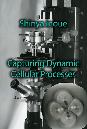
Schmidt had shown that mitotic spindles were birefringent. Inoué and Dan needed to harness this property to construct a polarizing microscope which would be able to clearly show the mitotic spindles over the course of cell division. For Inoué to obtain the contrast needed for observing the mitotic spindle, he would need to develop a microscope specially-designed for polarizing light, and not just add an element to a microscope designed for non-polarized light as Nicol had done. Optical microscopes include, from the bottom upwards, a light source, a condenser which comprises a lens and a diaphragm to control the aperture through which the light travels, a stage on which the slides are placed, an objective lens which focuses the light rays, and the ocular lens or eyepiece that focuses the image for the observing microscopist. Inoué’s painstaking efforts to construct a microscope to show the spindles through cell division resulted in what his colleagues christened, ‘the Shinya-scope’. We now call it ‘Shinya-scope 1’, as it was just the first of many.
- Shinya Inoué, Pathways of a Cell Biologist. Through Yet Another Eye. Springer: 2016
- Shinya Inoué, Collected Works Of Shinya Inoué: Microscopes, Living Cells, and Dynamic Molecules (With DVD-ROM). World Scientific Publishing Company; Har/Dvdr edition (July 18, 2008).

