Still Image
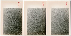
Three black and white photos of cells. The photos have been labelled along the side with times (2:40, 2:45, 2:50), and several cells in each have been marked with a red dot.
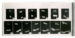
13 black and white photographs of different stages of chick limb bud development. The limb buds have been removed from the embryo and are placed on a black background. Stages 23 through 35 are pictured. Wing buds are pictured on top, leg buds on the bottom.
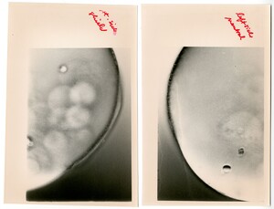
Two black and white photos of different sides of an embryo. The right photo is labelled "left side neutral" and the left photo is labelled "right side shield".
15 hand drawn sketches in pencil of exogastrule. Sketches are labelled in ink: 23 through 35b, and notes in ink accompany
13 hand drawn sketches in pencil of exogastrulae. Sketches labelled in ink: 13 through 26.
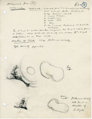
Hand drawn sketch of chick embryo (4.5 days incubation) and the development of the wing and limb buds. Also hand written notes about the stage of the embryo and the differences between it and other stages.
Crepidula embryos from a microscope slide made by Edwin Grant Conklin
Hand drawn sketches of early-stage chick limb bud development along with hand written notes about the embryo.
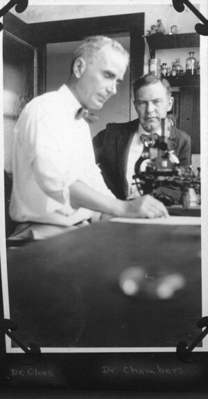
Dr. Clues and Dr. Chambers sitting at a lab bench with a microscope.
Dr. Parmenter standing outside in front of a building with his hands folded behind his back.
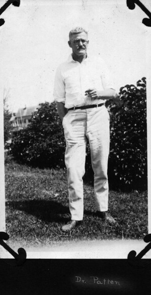
Patten standing outside in front of some bushes with one hand placed in his pocket
Plough standing in front of a wall of a building with a pipe held up in his right hand.

