Still Image
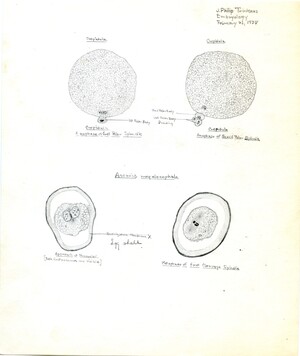
Labeled drawing by Trinkaus of Ascaris megalocephala, Embryology course, February 21, 1938
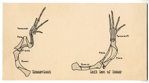
Hand drawn illustration of the outcome of chick limb bud transplantation
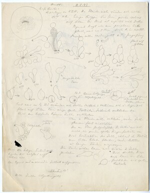
Hand drawn sketches pencil of cells and embryos along with with hand written notes
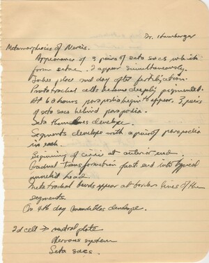
Notes from Viktor Hamburger's lecture. Trinkaus begins his notes with information on "Metamorphosis of Nereis", including information on the derivatives of the "2d cell"
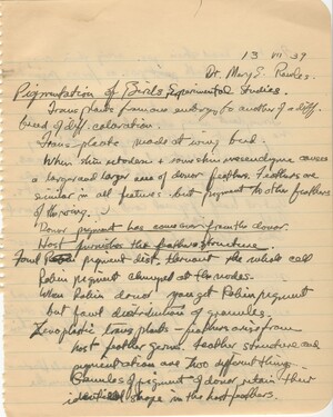
Notes from Mary Rawles' lecture. Trinkaus begins his notes with information on "Pigmentation of Birds, Experimental Studies" including information on transplantation experiments
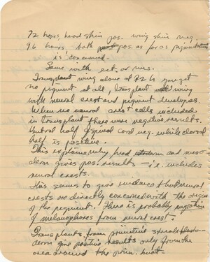
Notes from Mary Rawles' lecture. Trinkaus continues his notes from on transplantation experiments for bird pigmentation from the previous page. Trinkaus gives details about the experimental procedure and pays special attention to the role of the neural crest
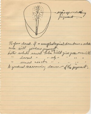
Notes from Mary Rawles' lecture. Trinkaus concludes his notes on pigmentation experiments in birds with a diagram of the neural tube with cells that produce pigment
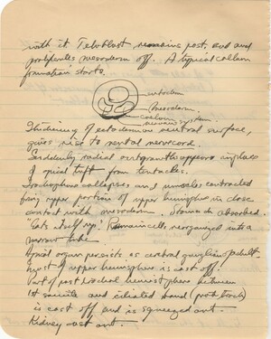
Notes from Viktor Hamburger's lecture. Trinkaus continues his notes on the metamorphosis of the trochophore with a diagram and information about how it changes during development
Notes from Viktor Hamburger's lecture. Trinkaus concludes his notes for this lecture with a final line about the metamorphosis of the trochophore
Notes from Roberts Rugh's lecture. Trinkaus continues his notes from the previous page on the effects of X Rays on amphibian eggs
Hand drawn sketches of Ambystoma (a genus of salamander) embryos, along with notes about their development
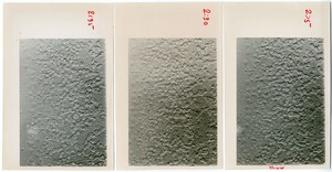
Three black and white photos of cells. The photos have been labelled along the side with times (2:15, 2:30, 2:35), and several cells in each have been marked with a red dot. The right image is labelled "shield" at the bottom, indicating the side of the embryo where the embryonic shield was forming.

