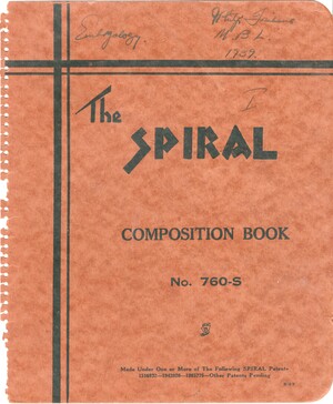
Trinkaus, John Phillip - Papers
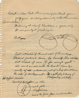
Notes from Hubert Goodrich's lecture. Trinkaus continues Periblast notes from page 8. Begins a new section on 'Gastrulation' with two diagrams of the 'embryonic shield', and a diagram and explanation of 'Kupffer's Vessicle'
Notes from Hubert Goodrich's lecture. Trinkaus has drawn four colored diagrams of 'Presumptive Regions': Trout, Fundulus, Dog fish, and Urodele
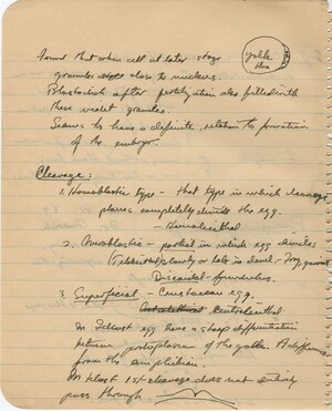
Notes from Hubert Goodrich's lecture. Trinkaus continues notes from page 5 on the movement of nuclear granules early in development, and then begins notes on 'Cleavage'
Notes from Oscar Schotte's lecture. Trinkaus continues his notes on the development of the egg with information on 'internal potencies', 'visibles structure', and 'Invisible structure', then begins a new section on 'External Factors" with a diagram of a two-cell embryo
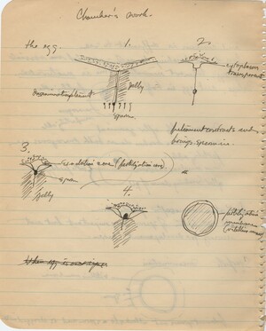
Notes from Oscar Schotte's lecture. Trinkaus draws 4 labeled diagrams of Chamber's work on fertilization in starfish
Notes from Oscar Schotte's lecture. Trinkaus takes notes on the "layers of the egg" with a diagram, then details the work of Chambers on the anatomy and structure of the echinoderm egg
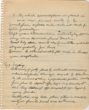
Notes from Viktor Hamburger's lecture. Trinkaus continues notes from page 57 and 58 on the copulation behavior of squid. He starts a new section on "Egg Laying"
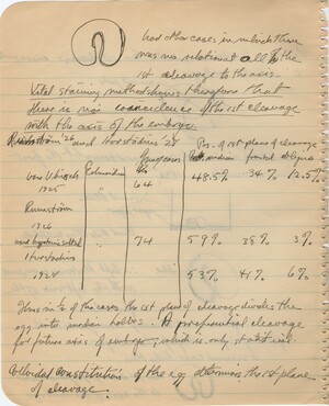
Notes from Oscar Schotte's lecture. Trinkaus continues his notes on the echinoderm egg notes about the position of the first cleavage relative to the axis of the embryo. He offers results of various researcher's studies and concludes that "Vital staining method shows therefore that there is no coincidence of the 1st cleavage with the axis of the embryo"
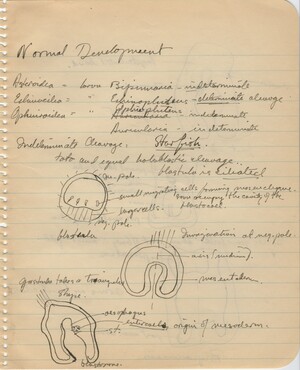
Notes from Oscar Schotte's lecture. Trinkaus begins notes on 'Normal Development' with information on Asteroidea, Echinoidea, Ophiuroidea, including 3 labeled diagrams
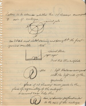
Notes from Oscar Schotte's lecture. Trinkaus continues his notes on the echinoderm egg with information about the position of first cleavage relative to the axis of the embryo, with four diagrams
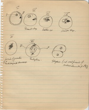
Notes from Oscar Schotte's lecture. Trinkaus draws 6 diagrams of fertilization and the first cell division.
Signed "J. Philip Trinkaus, M.B.L., 1939, Embryology, I"

