Specimen
Black and white photograph of net with horseshoe crab
Black and white photograph of a jar with a preserved fish
Black and white photograph of large jar with preserved sea stars
Black and white photograph of jars of sea star specimens
Black and white photograph of jar with preserved fish
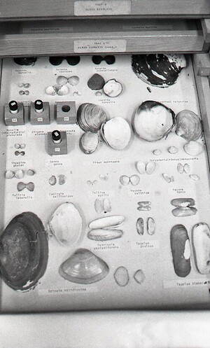
Black and white photograph of board with preseved shells
Black and white photograph of polarizing microscopy of the mitotic spindle of the endosperm. The two columns depict images obtained with different rotations of the compensator. The top row depicts the cell during metaphase, the bottom four rows depict, from the second row down to the fifth, progression through anaphase.
Polarizing microscopical images of crystals of Green Fluorescent Protein. This work, with Inoue conducted with Osamu Shimomura and others, has shown that it is possible for researchers to identify the orientation of GFP in cells, as well as its presence or absence.
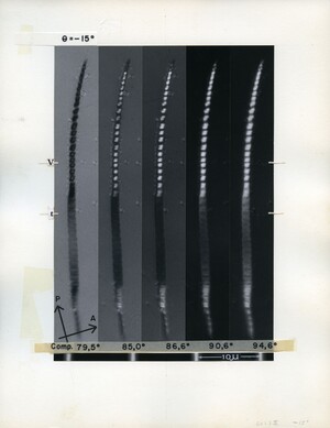
Polarizing microscopical image of cave cricket sperm. The patterns of birefringence in the upper half of the sperm displays the different regions of the DNA. The dark region below the middle of the sperm has been irradiated with UV. The five images were taken at different rotational angles of the compensator, which allow light of different polarizations to pass through.
Black and white photograph of microscopic image of sand dollar embryo. The image shows four cells with the birefringence of the spindle fibers contained within them. Whether the birefringence is bright or dark depends on the orientation of the spindle. The main object of polarized light microscopy is contrast, and this is evident in this picture. Shinya has written on the back of the photograph: 75e22I From 17A RCF-3.5 15sec@F/11 2mD


