Notes & Data
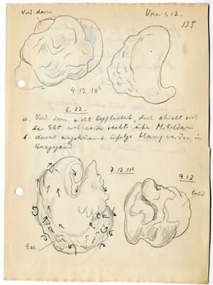
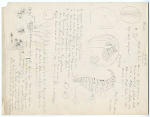
Hand drawn sketches in pencil and colored pencil of exogastrulae, along with hand written notes in pencil. One of the exogastrulae is covered in small arrows, indicating the movement of cells in different directions as the embryo expands.
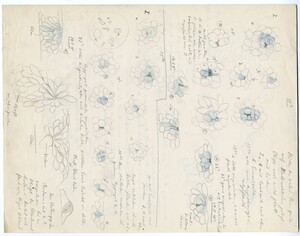
Hand drawn sketches of cells (some colored in blue) along with hand written notes (pencil)
3 micrographs and 2 hand drawn images of chick somite development. Top left shows a piece of a transverse section of a stage 10 chick embryo. Middle top and bottom shows a close up of the somite (transverse section). Right top and bottom are hand drawn images of cells in the somites.
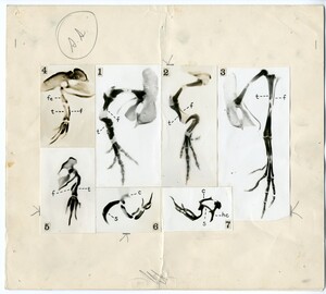
7 micrographs showing the results of chick limb transplantation
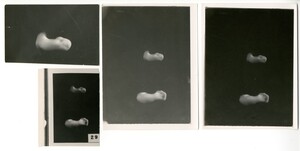
4 black and white photographs of chick limb bud development. Stage 29. The limb buds have been removed from the embryo and are placed on a black background. The image on the top left is of only a wing bud at stage 29.
Hand drawn sketches pencil of cells and embryos along with with hand written notes
10 black and white photographs of transverse sections through various stages of chick embryos. The bright white oval in the center of each section is the central canal of the developing chick's neural tube. The photographs are arranged so that dorsal is up.
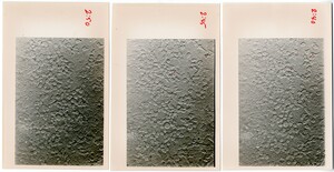
Three black and white photos of cells. The photos have been labelled along the side with times (2:40, 2:45, 2:50), and several cells in each have been marked with a red dot.
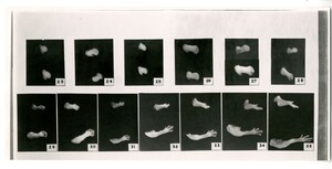
13 black and white photographs of different stages of chick limb bud development. The limb buds have been removed from the embryo and are placed on a black background. Stages 23 through 35 are pictured. Wing buds are pictured on top, leg buds on the bottom.
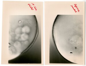
Two black and white photos of different sides of an embryo. The right photo is labelled "left side neutral" and the left photo is labelled "right side shield".
15 hand drawn sketches in pencil of exogastrule. Sketches are labelled in ink: 23 through 35b, and notes in ink accompany
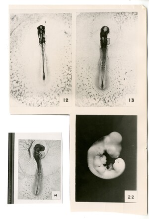
4 black and white photographs of different stages of chick development (whole embryos). Stages 12, 13, 14, and 22 are shown.

