Steinert
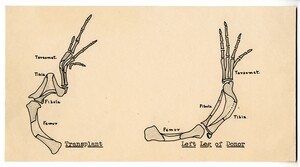
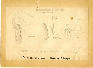
Hand drawn illustration of the outcome of chick limb bud transplantation
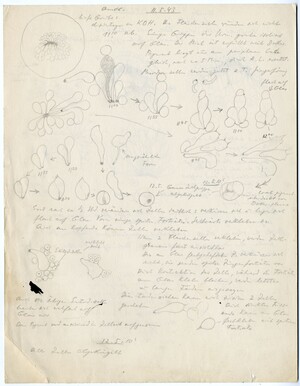
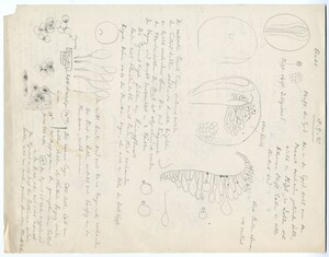
Hand drawn sketches pencil of cells and embryos along with with hand written notes
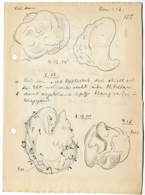
Hand drawn sketches in pencil and colored pencil of exogastrulae, along with hand written notes in pencil. One of the exogastrulae is covered in small arrows, indicating the movement of cells in different directions as the embryo expands.
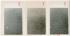
Three black and white photos of cells. The photos have been labelled along the side with times (2:15, 2:30, 2:35), and several cells in each have been marked with a red dot. The right image is labelled "shield" at the bottom, indicating the side of the embryo where the embryonic shield was forming.
4 black and white photographs of exogastrulae (appear to be amphibian). The photos have been pasted to a skeet of blue card stock and are labelled in ink.
Hand drawn sketches of Ambystoma (a genus of salamander) embryos, along with notes about their development
Hand drawn sketches in pencil and colored pencil of exogastrulae, along with hand written notes in pencil. Some of the exogastrulae are labelled with hours and constitute a time series. The colors indicate the movements of different cell groups.
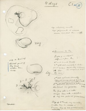
Hand drawn sketch of chick embryo (4 days incubation) and the development of the wing and limb buds. Also hand written notes about the stage of the embryo and the differences between it and other stages.
Hand drawn illustration showing how to transplant limb buds in a 36-somite stage (3 days of incubation) chick.
Hand drawn sketches pencil of cells and embryos along with with hand written notes
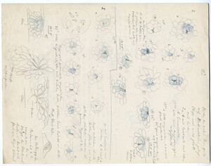
Hand drawn sketches of cells (some colored in blue) along with hand written notes (pencil)
3 micrographs and 2 hand drawn images of chick somite development. Top left shows a piece of a transverse section of a stage 10 chick embryo. Middle top and bottom shows a close up of the somite (transverse section). Right top and bottom are hand drawn images of cells in the somites.

