Shinya Inoue
Reconstruction of Shinya-scope 1 drawn from memory by Shinya Inoue in October 1992. Inoue had previously rested the parts of his polarizing microscope on books, but when the Emperior of Japan came to visit, he went to try and find a base to attach the parts of the microscope to. He found a discarded machine gun base, and attached the parts to it with string.
Color photograph of Shinya Inoue (left) with his mentor Katsuma Dan (right). Photograph taken by Y. Hiraiyoto and sent to Inoue.
Black and white photograph of polarizing microscopy of the mitotic spindle of the endosperm. The two columns depict images obtained with different rotations of the compensator. The top row depicts the cell during metaphase, the bottom four rows depict, from the second row down to the fifth, progression through anaphase.
Polarizing microscopical images of crystals of Green Fluorescent Protein. This work, with Inoue conducted with Osamu Shimomura and others, has shown that it is possible for researchers to identify the orientation of GFP in cells, as well as its presence or absence.
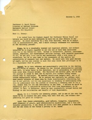
Letter of recommendation written by Kenneth Cooper to Marsh Tenney, as part of Inoue's ultimately successful application for a position at Dartmouth College.
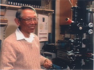
Color photograph of Shinya Inoue with a microscope at the Marine Biological Laboratory
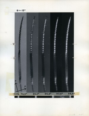
Polarizing microscopical image of cave cricket sperm. The patterns of birefringence in the upper half of the sperm displays the different regions of the DNA. The dark region below the middle of the sperm has been irradiated with UV. The five images were taken at different rotational angles of the compensator, which allow light of different polarizations to pass through.
Black and white photograph of Shinya Inoue in his laboratory with one of his microscopes.
Shinya-scope 3. Taken at the University of Washington. Photograph produced by University of Washington Department of Medical Photography, number 4054.
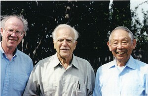
Photograph of three scientific generations. From left to right, Ted Salmon, Shinya's PhD student; Kenneth Cooper, Shinya's supervisor at Princeton; and Shinya Inoue.
Black and white photograph of microscopic image of sand dollar embryo. The image shows four cells with the birefringence of the spindle fibers contained within them. Whether the birefringence is bright or dark depends on the orientation of the spindle. The main object of polarized light microscopy is contrast, and this is evident in this picture. Shinya has written on the back of the photograph: 75e22I From 17A RCF-3.5 15sec@F/11 2mD


