photographs
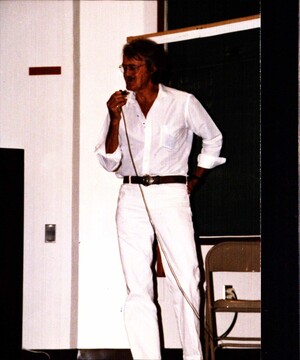
Photo of John Philip Trinkaus on August 12, 1988; description written on back possibly mentions Old Timers' Day
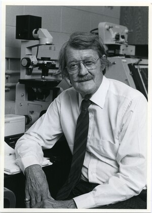
John P. Trinkaus wearing a long-sleeved white shirt, striped tie, and glasses. Trinkaus has a moustache and is seated in a laboratory, in front of a microsdope. He is leaning on a note pad and looking at the camera.
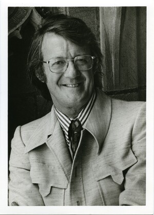
John P. Trinkaus is wearing a a striped shirt, light colored jacket, paisley tie, and glasses. Trinkaus is standing in front of a stone wall, looking at the camera and smiling.
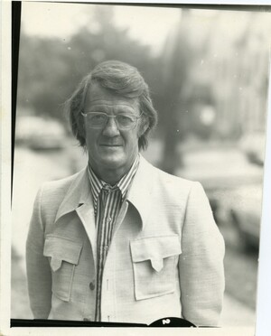
John P. Trinkaus wearing a striped shirt, light colored jacket, and glasses. Trinkaus is outside, and looking directly at the camera.
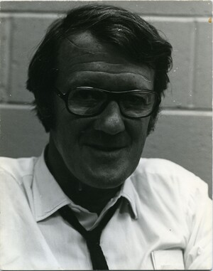
A headshot of John P. Trinkaus. Trinkaus is wearing a white shirt, dark tie, and glasses. He is looking at the camera.
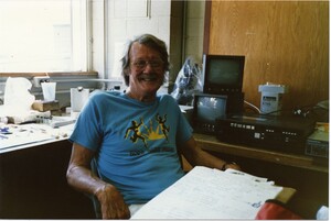
John P. Trinkaus in the laboratory at the MBL. Trinkaus is seated in front of video equiptment, and is smiling at the camera.
John P. Trinkaus (left) standing next to Steve Zottoli (right) in their shared laboratory at the MBL
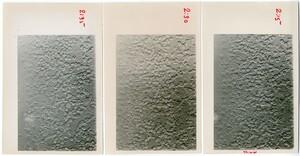
Three black and white photos of cells. The photos have been labelled along the side with times (2:15, 2:30, 2:35), and several cells in each have been marked with a red dot. The right image is labelled "shield" at the bottom, indicating the side of the embryo where the embryonic shield was forming.
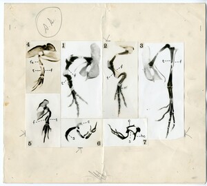
7 micrographs showing the results of chick limb transplantation
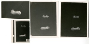
4 black and white photographs of chick limb bud development. Stage 29. The limb buds have been removed from the embryo and are placed on a black background. The image on the top left is of only a wing bud at stage 29.
10 black and white photographs of transverse sections through various stages of chick embryos. The bright white oval in the center of each section is the central canal of the developing chick's neural tube. The photographs are arranged so that dorsal is up.
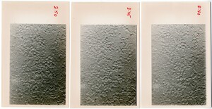
Three black and white photos of cells. The photos have been labelled along the side with times (2:40, 2:45, 2:50), and several cells in each have been marked with a red dot.

